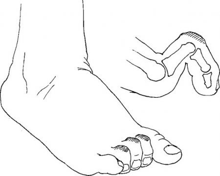
DIGITUS MALLEUS/HAMMER TOE
Deformation of the toes in the joint closer to the tiptoe with bent phalange in the form of a hammer. It is most common to develop on the second and third finger. It creates callus on the tiptoe along the top of the nail. Due to deformation, shoe often forms friction on the skin resulting in formation of callus. It can be treated with pedicure, but despite pedicure, it will recreate as long as there is no deformation. The most common cause of creation is the collapse of the transverse arch.
It is successfully treated with surgery.

DIGITUS FLEXUS/CLAW TOE
Deformation of the toes joints that are bent towards the foot. On crumpled toe over time above joint first appears skin redness due to the pressure of the shoe and then after that skin becomes hard as a sign of creating a corn. Sometimes it comes to a gradual dislocations on the toe base which leads to increased pressure on joint from the side of the feet and there may appear hard skin. Claw toe can be straighten only with surgery.


HALLUX RIGIDUS
“Stiff big toe” is characterized by arthrotic changes of first metatarsophalangeal joint accompanied by pain and dorsal flexion limitation. It is a consequence of ossification disorders in head of first metatarsal bone in youth. In adults it is caused by degenerative changes with dominated stiffness of the big toe basal joint.
Big toe is usually outstretched, dorsal flexion is reduced and plantar preserved. It is characterized by the classical signs of osteoarthritis such as narrowing of the joint cavity and osteophytes. Big toe is thickened in the first metatarsophalangeal joint due to atrhrotic changes. People with hallux rigidus due to pain often walk on the outer edge of the foot that can be seen on their shoes.


HALLUX VALGUS
Hallux valgus is the most common foot deformity. It is a distortion of the big toe to the side creating painful bony protrusions on the inner side of the root of the big toe.
Congenital of during life increased mobility of the first metatarsal bone is one of the causes for developing hallux valgus. In this case, first metatarsal bone moves away under pressure from other bones and raises abandoning its position as the lowest places bone in the front part of the foot. In that way it collapses the anterior arch that is composed of the heads of the metatarsal bones.


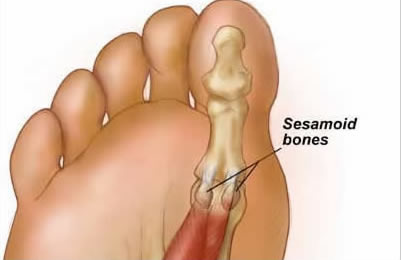
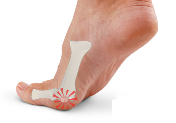
SESAMOIDITIS
The sesamoids are located in areas where tendons are crossing over joints. In foot they are located under head of fist metatarsal bone. Their function is to protect the tendons and to increase mechanical effect. Sesamoiditis indicates inflammation of the sesamoids. The most common symptoms are: pain under head of first metatarsal bone that gets worse while walking and wearing tighten shoes, redness, swelling, numbness of the front part of the foot. Running, long walking and sports that often include jumping on the frontal part of the foot such as tennis and basketball can cause fracture of sesamoids.
MORTON’S NEUROMA
Morton’s neuroma is caused by pressure on a nerve that goes through a metatarsal tunnel and it is interesting that it was first described by Morton back in 1876. Most often affected nerve is the one
that passes through III. metatarsal tunnel (space between heads of III. and IV. metatarsal bones) and much rather nerves that pass through II. and IV. metatarsal tunnel.
Morton’s metatarsalgia is usually associated with wearing high heel shoes and shoes that are too narrow in the front. Also, gestures that lead to excessive stretching of the toes, so called hyperextension of the toes (e.g. sprinting , running uphill and some professions that require longer squat), lead to a narrowing of the metatarsal tunnels and constantly reducing space for nerves causing their swelling and thus nerves become thicker and more prone to a further damage.


FLAT FEET
Flat feet is defined as a partial or complete loss of medial longitudinal arch which is present after the end of growth and development. Flat feet in adults may be an incidental finding on clinical examination, but it may be present as a symptomatic condition in range from minor issues to serious disorders that can significantly impair quality of life.
Causes of flat feet in adulthood are diverse and they include disorders of posterior tibial tendon, coalescence of tarsal bones, different post-traumatic and inflammatory conditions (rheumatoid arthritis, seronegative spondyloarthropathies) and neuromuscular pathologies such as diabetes and Charcot foot. Cramps, fatigue and pain occur in the muscles of the foot and lower leg.


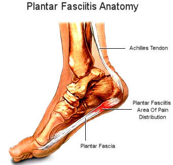
PLANTAR FASCIITIS
Plantar fasciitis is manifested as a pain at the bottom of the calcaneus and it occurs as a result of accumulated micropitting causing deterioration (degeneration) of collagen fibers in the fascia on the calcaneus and it belongs to a group of injuries know as overuse injuries.
Plantar fascia is placed under the foot skin and it is separated from her with the layer of the fat (fat at the heel is thicker more than 2 cm) and has three parts: middle, medial and lateral. The middle part is the most important. That is a strong triangular tendon plate that starts at the bulge of calcaneus and it is directed forward.
The main symptom is pain at the starting point of plantar fascia on calcaneus. Sometimes, pain can be spread inside of the foot while the swelling is extremely rare. There is characteristic appearance of very strong pain in the morning when getting up from bed which disappears after a dozen steps.

ACHILLES TENDINOSIS
Achilles tendon is the final part of a three headed muscle of the lower leg (lat. triceps surae) and it catches on the bottom half of the calcaneus back. This is the strongest tendon in the human body with of length 5 – 6 cm and and thickness 5 – 6 mm.
Achilles tendinosis is associated with influence of the force on Achilles tendon while walking and/or running which is increased due to influence of some predisposing factors – anatomical feet deviations (e.g. flat feet), excessive pronation while walking and/or running, disproportion between strength and flexibility of the muscles that form Achilles tendon and age (age decreases the tendon elasticity). The main symptom is pain localized 2 – 5 cm above attaching point on calcaneus which is characteristically related to the activity because it appears at the beginning, decreases during and increases after activity. Another characteristic symptom is an appearance of morning pain and stiffness in the talocrural joint which disappears after few steps. In some cases, especially in those who have occurred sudden, patients complain about creaking along the tendon.


CHRONIC ANKLE INSTABILITY

Most ankle sprains are inversion type. When a patient twist his ankle, the bottom of the foot is turned toward second leg and the outside of the foot makes contact with the ground. In rare cases, foot may turn in opposite direction. These excessive and sudden movements will pull the ligament
and act with great force. Although ligaments are strong, those forces are strong enough to damage the ligament. The ligaments will extend, or partly or completely rupture, and their grip can be pulled out from the bone or even tear off a piece of the bone.
After the injury, there is swelling and pain outside of the ankle and foot. There will be bruising and skin discoloration to blue-green color. The pain is spreading through the bone on the outside of the ankle across ligaments and tendons in the ankle. In this condition, patients will be constantly on the alert during a walk because of their feeling of twisting it again. This can be described as unstable ankle that can often lead to a chronic pain.