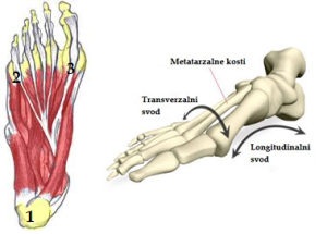
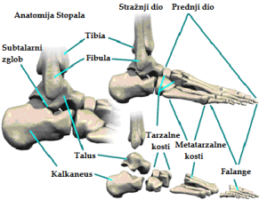
During a walk, body weight is transferred from calcaneus to the first and fifth metatarsal bone making a reference points of the foot. Those three points are connected by ligaments, muscles and bones forming the medial and lateral longitudinal arch and the anterior and posterior transverse arch. Medial longitudinal arch starts from tuber calcanei, continues over talus, navicular, medial cuneiform bone along metatarsal bone to its head. The point of the medial longitudinal arch, which is the most lifted from the floor (15 – 20 mm), is the lower edge of the navicular bone. Lateral longitudinal arch is starts from tuber calcanei across cuboid along fifth metatarsal bone to its head. The highest point in lateral longitudinal arch is cuboid bone which is lifted from the base 3 to 5 mm. Anterior transverse arch connects the front reference points of the foot – head of first and fifth metatarsal bones. The highest point of the anterior transverse arch is head of the second metatarsal bone in adults and due to the inversion of the foot, head of the first metatarsal bone in children. Posterior transverse arch is located in area of the three cuneiform bones and cuboid bone.
 There are many tendons and ligaments that form foot arches among the most important are calcaneonavicular ligament, long plantar ligament and plantar aponeurosis. Also, all muscles of the lower leg (except muscle triceps surae) and foot are included in forming the arches. Muscles are the only active arch holders while bones, ligaments and tendons support passively.
